Research Highlight · Neuro-Oncology Advances
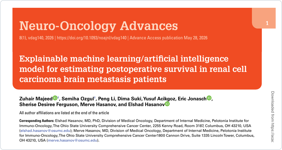
Explainable ML/AI model for postoperative survival in RCC brain metastasis
This article was published in Neuro-Oncology Advances and focuses on interpretable postoperative survival modeling for renal cell carcinoma brain metastasis.
Overview
This study develops an explainable ML/AI model for estimating postoperative overall survival after surgical resection of renal cell carcinoma brain metastases. The work was published in Neuro-Oncology Advances and focuses on a clinically challenging setting where outcome prediction can support risk stratification, surgical decision-making, and translational research.
The central idea is that survival modeling should be useful not only as a prediction task, but also as a way to organize clinical and radiologic signals into interpretable risk patterns. In other words, the goal is not simply to produce a score; the goal is to make postoperative risk estimation more transparent and clinically meaningful.
Clinical Motivation
Brain metastasis from renal cell carcinoma is a difficult clinical scenario. Patients may present with very different systemic disease status, brain lesion burden, symptoms, and surgical context, and these factors can interact in ways that are hard to summarize with a single traditional staging system.
Existing prognostic tools are helpful, but many are either not specific to RCC brain metastasis or do not fully incorporate detailed surgical, radiologic, and clinical variables. This matters because surgery for brain metastasis is not a purely technical decision: the expected survival horizon, neurologic symptom burden, extracranial disease status, and postoperative management plan all shape whether surgery is likely to benefit an individual patient.
For translational oncology, this creates a practical need for models that can estimate risk while still allowing researchers and clinicians to understand which factors contribute to the prediction.
Cohort and Modeling
The study retrospectively analyzed patients with histologically confirmed RCC and radiographically or pathologically confirmed brain metastases who underwent neurosurgical resection. After excluding cases with missing information needed for model development, the final modeling cohort included 253 patients.
The primary endpoint was overall survival after brain metastasis resection. Multiple clinical and radiologic variables were evaluated, and several survival modeling strategies were compared. The best-performing final approach combined XGBoostCox with a plsRcox-based model, aiming to capture non-linear prognostic patterns while still supporting model interpretation.
Rather than presenting machine learning as a generic black-box exercise, the analysis was framed around a focused neuro-oncology question: can detailed perioperative data improve individualized postoperative survival estimation for RCC brain metastasis patients?
Why Explainability Matters
In high-stakes clinical research, model transparency is important. A survival model may perform well statistically, but if the risk score cannot be understood or audited, it is harder to trust and harder to translate into follow-up studies.
To address that issue, the study used SHAP analysis to estimate how individual variables contributed to model predictions. This allowed the model to be inspected at both the population level and the patient level. The most influential factors included extracranial disease status, number of brain metastases, preoperative symptoms, and age at surgical resection.
This interpretability layer is central to the project. It helps move the model from a prediction-only tool toward a clinically inspectable framework for thinking about postoperative risk.
What We Found
The final model achieved a test concordance index of 0.59. Time-dependent AUCs at 1, 2, and 5 years were 0.61, 0.64, and 0.69 in the test cohort. These values reflect the difficulty of the clinical problem: survival after brain metastasis resection is influenced by many interacting factors, and the available cohort is clinically heterogeneous.
Importantly, the model-derived risk groups separated postoperative survival in the test cohort. Using a cutoff selected from the training cohort, the high-risk group had significantly worse survival than the low-risk group, with a hazard ratio of 2.06.
The result is useful because it combines three pieces that are often separated: survival modeling, model explainability, and clinical translation. The model does not merely rank patients by risk; it also provides a way to examine which features are driving that risk estimate.
Clinical Translation
A major translational component of the study is the online survival calculator built from the final model. The calculator is intended to make individualized postoperative risk assessment more accessible and to support future clinical discussion, validation, and refinement.
For a personal research portfolio, this project is a good example of how machine learning can be used in oncology without losing the clinical question. The computational part matters, but the motivating problem remains patient-centered: how can we better estimate postoperative prognosis for a high-risk metastatic population?
Key Takeaways
- The project applies ML/AI survival modeling to a focused renal cell carcinoma brain metastasis resection problem.
- The final model combines XGBoostCox and plsRcox, with performance evaluated using C-index and time-dependent AUC.
- SHAP analysis highlights clinically interpretable drivers of postoperative risk, including extracranial disease status and brain metastasis burden.
- The work includes an online calculator, making the model more accessible for translational use and future validation.